
A Cutting-Edge Microscope for a New Approach in Neuroscience
Enabling Significant Advances in the Treatment of Cranial and Brain Disorders at Hôpital Foch
As one of the centers of expertise at Hôpital Foch, the neurosurgery department manages all surgical conditions related to the brain and spinal cord.
THE PROJECT
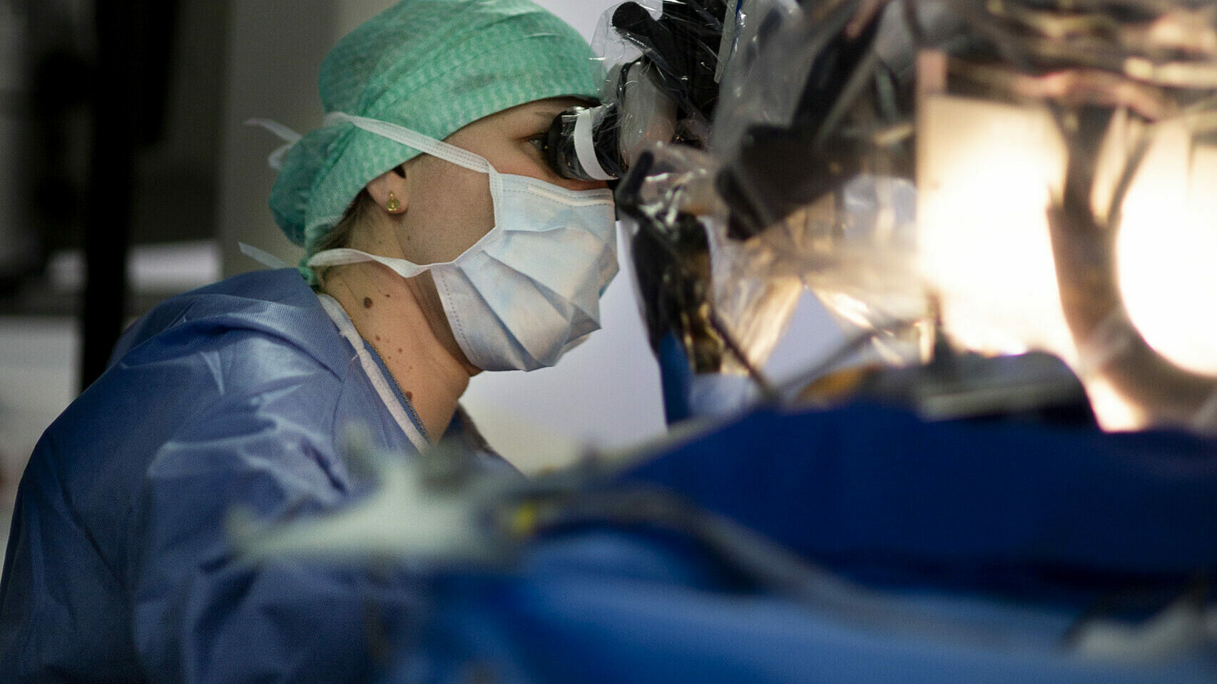
The acquisition of a surgical microscope with a fluorescence module marks a major advancement in skull base surgery, providing significant benefits in terms of precision, safety, and customization of surgical procedures. Investing in this advanced technology helps Hôpital Foch remain at the forefront of medical innovation, directly improving the quality of patient care.
Integrating a surgical microscope with a fluorescence module into modern surgical practices represents a groundbreaking technique for treating complex skull base conditions, such as atypical and recurrent meningiomas, as well as sinonasal cancers. This technology offers undeniable advantages, improving patient outcomes by enabling more precise and safer tumor removal.
Fluorescence enhances visualization of tumor tissue, allowing a clear distinction between malignant and healthy tissues. This increased precision is crucial for achieving complete resection, especially in cases of recurrences or meningiomas located in difficult-to-access areas of the skull base. The ability to accurately detect and remove tumor tissue can significantly reduce recurrence rates, a major benefit for patients who have already undergone multiple surgical and radiation treatments.
Skull base surgeries are notoriously complex due to the proximity of critical neurovascular structures. Fluorescence guidance in such cases allows for better identification and preservation of essential structures.
BENEFITS FOR PATIENTS
✔ Enhanced surgical precision
Fluorescence enables surgeons to more accurately distinguish malignant tissues from healthy ones.
✔ Significant reduction in recurrence rates
A more precise tumor resection reduces the risk of recurrence, improving long-term recovery prospects.
✔ Improved post-operative recovery
A more targeted procedure allows for faster healing and reduces postoperative complications.
✔ Advancement of research on cranial pathologies
This advanced equipment also serves as a valuable tool for training surgeons and conducting clinical research on surgical techniques.
Professor Damien Bresson, Head of the Neurosurgery Department, has been a pioneer in the development of innovative surgical techniques in France. His expertise in skull base surgery is complemented by clinical experience and participation in groundbreaking research.
🗣 “By choosing to support our project, you help Hôpital Foch integrate the latest technological innovations into our surgical practices, with the goal of offering our patients more precise, effective, and safer treatments. The adoption of the fluorescence-equipped microscope demonstrates our commitment to staying at the forefront of medical progress and continuously improving patient care. Thank you for your support.”
Glossary
🔹 Neurosurgery: A specialized field of the neuroscience department, focusing on surgical treatment of the central and peripheral nervous system, as well as spinal disorders.
🔹 Fluorescence: A light emission caused by the excitation of electrons in a molecule or atom, usually through photon absorption.
🔹 Resection: A surgical procedure that involves cutting and removing a portion of an organ or tissue.
The acquisition of a surgical microscope with a fluorescence module marks a major breakthrough in skull base surgery.